Blog
My New Glasses Aren't Working for Me
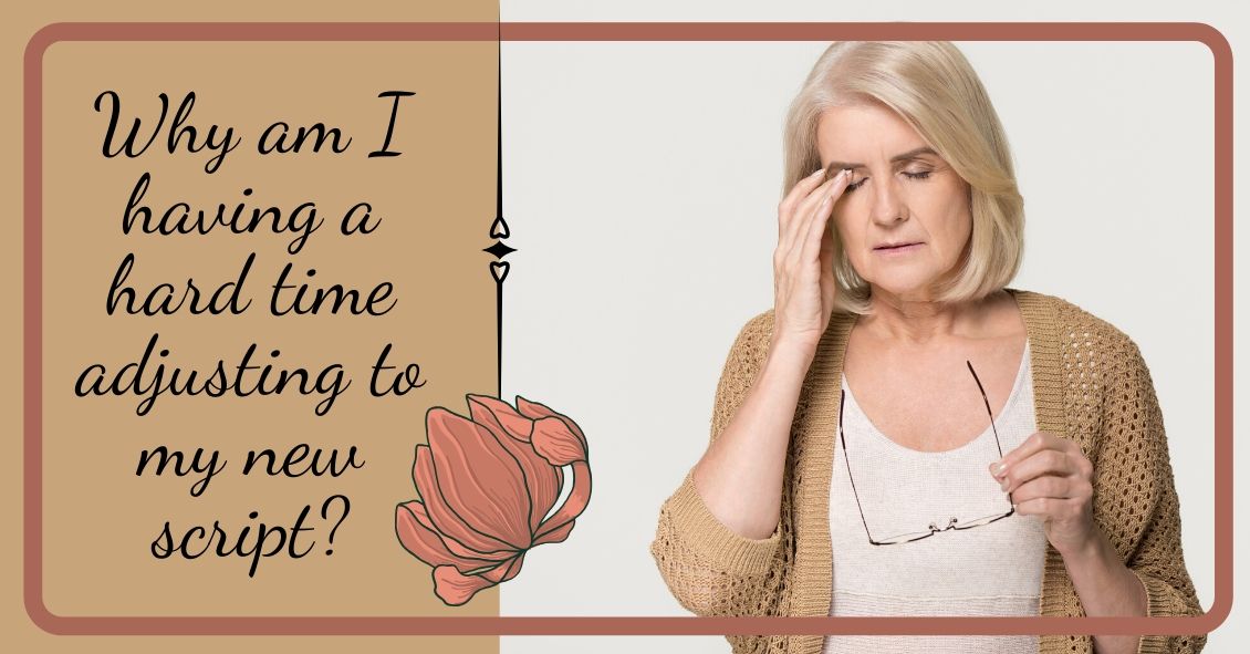
Eye doctors typically pride themselves on being able to improve someone’s vision through glasses or contact lens prescriptions. Whether it’s a first-time glasses wearer, or someone having either a small or large change in their prescription, we like to aim for that goal of 20/20 vision.
Despite our best efforts, however, correcting vision to 20/20 is not always a positive outcome for the patient. Whether someone will be able to tolerate their new prescription is based on something called...
The Wonderful World of Dailies
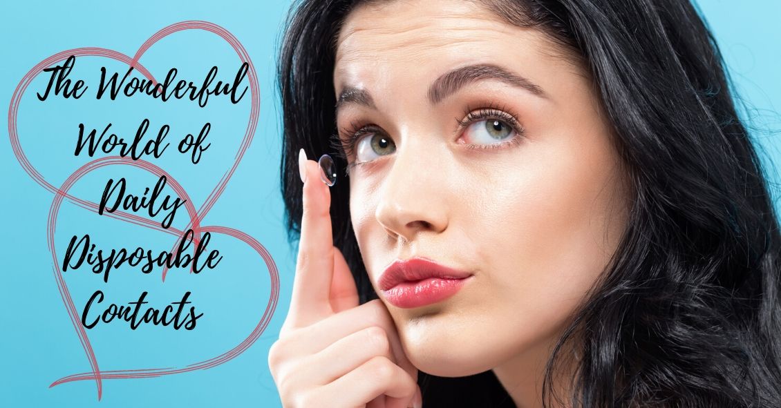
When soft contact lenses first came on the scene, the ocular community went wild.
People no longer had to put up with the initial discomfort of hard lenses, and a more frequent replacement schedule surely meant better overall health for the eye, right?
In many cases this was so. The first soft lenses were made of a material called HEMA, a plastic-like polymer that made the lenses very soft and comfortable. The downside to this material was that it didn’t allow very much oxygen to the...
6 Reasons Sunglasses Are Essential
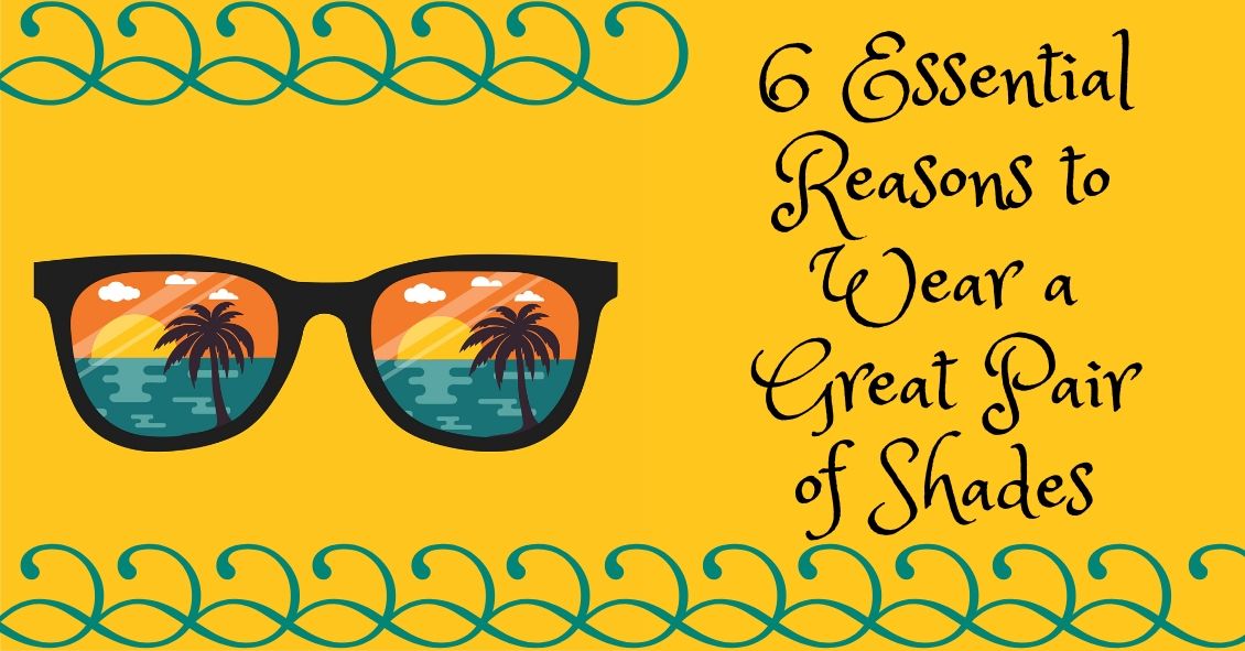
Sunglasses are more than just a fashion statement - they’re important protection from the hazards of UV light.
If you wear sunglasses mostly for fashion that’s great--just make sure the lenses block UVA and UVB rays.
And if you don’t wear sunglasses, it’s time to start.
Here are your top 6 reasons for wearing sunglasses:
Preventing Skin Cancer
One huge way that sunglasses provide a medical benefit is in the prevention of skin cancer on your eyelids. UV light exposure from the...